
Shoulder Muscles Anatomy, Exercise, Name List:
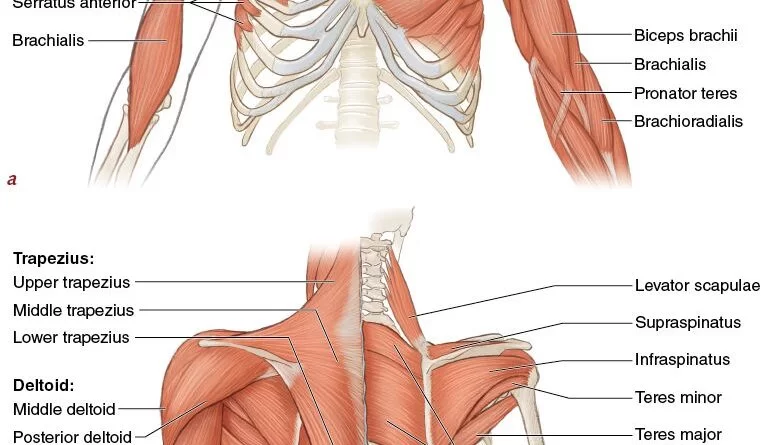
The shoulder muscles play a vital role in the complex mechanics of the shoulder joint. Shoulder girdle movements are supported and caused by the shoulder muscles. They link the appendicular skeleton of the upper limb to the axial skeleton of the trunk. Four of them are on the front of the shoulder, while the others are on the back and sides of the shoulder.
- The shoulder complex is composed of the clavicle, scapula, and humerus bone
- It is a combination of four joints, the Glenohumeral Joint, the Acromioclavicular Joint, and the Sternoclavicular Joint, and a “floating joint”, known as the Scapulothoracic joint.
- The shoulder joint also known as the glenohumeral joint is a ball and socket joint.
- The muscles of the shoulder have a wide range of functions, including abduction, adduction, flexion, extension, and internal and external rotation.
- the central bony structure of the shoulder is the scapula, where all of the muscles interact.
- The muscles of the shoulder play a critical role in providing stability to the shoulder joint.
- The primary muscle group that supports the shoulder joint is the rotator cuff muscles. There are four rotator cuff muscles includes supraspinatus, infraspinatus, teres minor, and subscapularis.
- Other muscles include:
- supraspinatus
- infraspinatus
- teres minor
- subscapularis
- Teres major
- Deltoid
- Serratus anterior
- Subclavius
- Pectoralis minor
- Pectoralis major
- Sternocleidomastoid
- Levator scapulae
- Triceps
- Biceps brachii
- Coracobrachialis
- Rhomboid major
- Rhomboid minor
- Trapezius muscle
- Latissimus dorsi
Intrinsic Muscle
- Known as the scapulohumeral muscular group, are deeper muscles that originate from the scapula and /or the clavicle and insert into the humerus.
- Supraspinatus
- Infraspinatus
- Subscapularis
- Teres minor
Extrinsic Muscle
- Extrinsic muscles are larger, more superficial muscles that originate in the thorax and attach to the bones of the shoulder complex.
- Latissimus dorsi
- Teres major
- Pectoralis major
- Pectoralis minor
Accessory Muscle
- There are also other muscles that act as secondary movers on the shoulder joint known as accessory muscles.
- They are generally in the pectoral area or in the upper arm region.
- Biceps brachii
- Triceps brachii
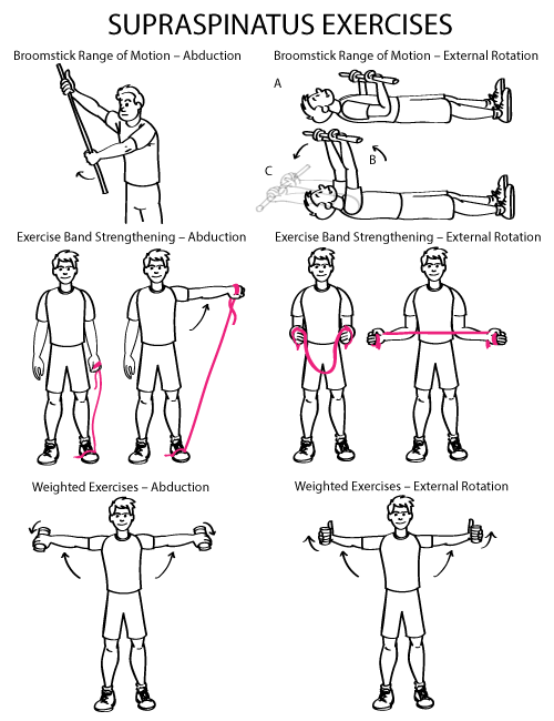
Supraspinatus
- Supraspinatus is the smallest of the 4 muscles which comprise the Rotator Cuff of the shoulder joint specifically in the supraspinatus fossa.
- It travels underneath the acromion.
- Origin :
- The supraspinatus muscle originates from the Supraspinatus fossa of the scapula.
- A shallow depression in the body of the scapula above its spine.
- Insertion :
- Greater tuberosity of the humerus, superior facet.
- Nerve Supply :
- Suprascapular Nerve, C5 & 6, superior trunk of the brachial plexus.
- Blood Supply :
- Suprascapular Artery.
- Action :
- It abducts the arm from 0 to 15 degrees, when it is the main agonist, then assists the deltoid to produce abduction beyond this range up to 90 degrees.
- It also, act as a shoulder stabilizer.
- Test for Supraspinatus :
- Empty Can Test: To test for supraspinatus impingement or integrity of the supraspinatus muscle and tendon.
- Patient’s position: Sitting or Standing.
- Therapist’s position: On the side to be tested and one of the therapist’s hands stabilizes the shoulder girdle
- Procedure: The arm to be tested is moved into 90 degrees of abduction in the plane of the scapula, 30 degrees of forward flexion, and full internal rotation with the thumb pointing down as if emptying a beverage can. The examiner’s other hand applies downward pressure on the superior aspect of the distal forearm and the patient resists.
- The Empty Can Test is considered positive if there is significant pain and/or weakness.
Infraspinatus
- A thick, triangular muscle one of the 4 muscles which comprise the Rotator Cuff of the shoulder.
- Origin :
- The infraspinatus fossa of scapula
- Insertion :
- The posterior aspect of the greater tuberosity of the humerus, and the capsule of the shoulder joint.
- Nerve Supply :
- Suprascapular Nerve (C5 & C6)
- Blood Supply :
- Suprascapular and circumflex scapular arteries.
- Action :
- Infraspinatus is the main external rotator of the shoulder joint.
- It assists in producing shoulder extension.
- Clinical Relevance :
Atrophy in the supraspinatus and infraspinatus muscles usually indicates compression on the suprascapular never near the scapular notch, which can be seen in overhead athletes and SLAP lesions. - While isolated weakness or atrophy in the infraspinatus muscle can be seen in suprascapular nerve compression in the spinoglenoid notch, which is due to a ganglion cyst.
Teres Minor
- Teres Minor is a narrow muscle that lies below the infraspinatus, above Teres major and triceps brachii, and deep to the deltoid.
- It is one of the four muscles which comprise the Rotator Cuff.
- Origin :
- The upper two-thirds of the lateral border of the scapula.
- Insertion :
- The upper fibers end in a tendon that inserts into the inferior facet of the greater tubercle of the humerus. The lower fibers insert into the humerus directly below the inferior facet of the greater tubercle of the humerus.
- Nerve Supply :
- The axillary nerve(C5, C6)
- Blood Supply :
- The circumflex scapular artery and the posterior circumflex humeral artery.
- Action :
- Teres Minor, along with Infraspinatus, primarily produces external rotation of the shoulder joint.
- It assists in the adduction and extension of the shoulder.
- When the humerus is stabilized, abducts the inferior angle of the scapula.
- Clinical relation :
- Hornblower’s sign: can be used to test the teres minor for tear.
- The patient’s arm should be placed at 90 degrees of scaption with the elbow flexed to 90 degrees.
- The patient will then externally rotate against resistance, trying to make a “field goal” sign.
- If the test is positive, the patient cannot externally rotate the shoulder, indicating teres minor pathology.
Subscapularis
- The subscapularis muscle is a large triangular-shaped muscle that originates from the subscapular fossa.
- The term “subscapularis” means under the scapula.
- It is part of the four rotator cuff muscles.
- The subscapularis is the largest and strongest muscle of the rotator cuff.
- Origin :
- Medial two-thirds of the subscapular fossa.
- Insertion :
- The fibres form a tendon that inserts into the lesser tuberosity of the humerus and the front of the shoulder joint capsule.
- Nerve Supply :
- Upper and lower subscapular nerves (C5-C6)
- Blood Supply :
- Subscapular artery
- Action :
- The primary function is the internal rotation of the humerus. It helps in shoulder adduction and extension.
- Clinical relation :
- The subscapularis can tear from overuse, trauma, or age-related conditions.
- Tears can be small or can go through most of the muscle.
- Symptoms include pain that gets worse at night, shoulder or arm weakness, and pain that gets worse when you lift your arm.
Teres Major
- Teres major is a small muscle that runs along the lateral border of the scapula.
- It forms the inferior border of both the triangular space and quadrangular space.
- It’s sometimes called “lat’s little helper” because of its synergistic action with the latissimus dorsi.
- Origin :
- The posterior surface of the inferior angle of the scapula.
- Insertion :
- Medial lip of intertubercular suclus of humerus.
- Nerve Supply :
- Lower subscapular nerve
- Blood Supply :
- Circumflex scapular artery
- Actions :
- Adducts and internally rotates the arm.
- Clinical relation :
- The teres major is a common muscle to the development of trigger points which prone to develop myofascial pain syndrome.
- Causes include poor stretching before physical activities and microtrauma through chronic inappropriate straining.
- Symptoms include local pain, which may radiate to the lateral shoulder, and difficulties in abducting and elevating the arm.
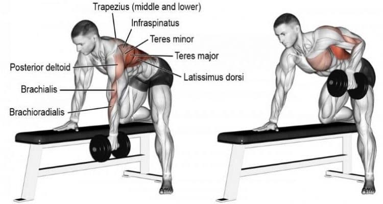
Deltoid
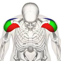
- The Deltoid muscle is a large triangular-shaped muscle that gives the shoulder its rounded contour.
- It is named after the Greek letter delta, which is shaped like an equilateral triangle.
- It comprises 3 distinct portions each of which produces a different movement of the glenohumeral joint, commonly named the anterior, medial, and posterior heads.
- Origin :
- Anterior deltoid :
- Anterior Surface of the lateral third of the Clavicle
- Medial deltoid :
- Acromion Process, Superior Surface.
- Posterior deltoid :
- The spine of the Scapula, Posterior Border.
- Insertion :
- Fibres from all heads converge to insert into the deltoid tuberosity on the humerus.
- Nerve Supply :
- Axillary Nerve, C5 &C6
- Blood Supply :
- The posterior circumflex humeral artery.
- Actions :
- All heads of the deltoid work together to produce abduction of the Shoulder Joint.
- While each individual head produces the following:
- Anterior deltoid :
- Flexes, abducts, medially rotates, and horizontally flexes the arm at the shoulder joint.
- Lateral deltoid :
- Abduction of the arm beyond the initial 15 degree
- Posterior deltoid :
- Extends, abducts, laterally rotates, and horizontally extends the arm at the shoulder joint.
- Clinical relation :
1)The axillary nerve can be damaged during dislocation of the shoulder joint or compressed during incorrect use of crutches. - Symptoms include atrophy of the deltoid muscle, resulting in weakness and a loss of muscle tone, making the shoulder look flattened rather than rounded.
- There may be a loss of sensation in the skin overlying the deltoid muscle.
- 2)The location of the axillary nerve is also important during intramuscular injections given in the deltoid muscle and during surgical approaches to the shoulder to avoid injuring the nerve.
Serratus Anterior
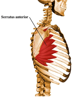
- The serratus anterior muscle is a fan-shaped muscle at the lateral wall of the thorax.
- Its main part lies deep under the scapula and the pectoral muscles.
- It is easy to palpate between the pectoralis major and latissimus dorsi muscles.
- Origin :
- The top lateral surface of the eight or nine upper ribs.
- Insertion :
- It inserts exactly at the front border of the scapula. The muscle is divided into three parts:
- 1)Superior: 1st to 2nd rib, superior angle of the scapula.
- 2)Middle : 2nd to 3rd rib , medial border of scapula
- 3)Inferior: 4th to 9th rib, medial border, and inferior angle of the scapula. It is the most powerful and prominent part.
- Nerve :
- The long thoracic nerve (C5 to C7)
- Blood supply :
- Lateral thoracic artery, the superior thoracic artery, and the thoracodorsal artery.
- Action :
- The main actions are protraction and upward rotation of the scapulothoracic joint, moving the scapula forward across the thoracic wall.
- It also keeps the medial border and inferior angle of the scapula close to the thoracic wall.
- Clinical relation :
- Long thoracic nerve damage may result from nerve compression due to anesthesia or Saturday night palsy, which is a radial neuropathy due to falling asleep with one’s arm hanging over the armrest of a chair.
- Heavy load bearing may also entrap the nerve, resulting in atrophy of the serratus anterior and subsequent winging of the scapula.
Subclavius
- The subclavius muscle is a short, triangular muscle of the thoracic wall that lies underneath the clavicle.
- Origin :
- Subclavius originates from the first rib.
- Insertion :
- It courses laterally to insert on the inferior surface of the middle third of the clavicle.
- Nerve supply :
- Subclavian nerve
- Blood supply :
- Clavicular branch of the thoracoacromial artery and the suprascapular artery.
- Action :
- Depression of the clavicle and stabilize the clavicle during movement of the shoulder girdle.
- Clinical relation :
- The blood vessels and nerves running behind the subclavius muscle can sometimes become entrapped between the clavicle and first rib, inside the costoclavicular space.
- This is known as a costoclavicular syndrome.
Pectoralis Major
- The pectoralis major is large and fan-shaped most superficial muscle in the pectoral region.
- It is composed of two heads: 1) clavicular head, 2) sternal head
- Origin :
- 1) clavicular head: originates from the anterior surface of the medial half of the clavicle.
- 2) sternal head: largest one,1) Anterior surface of the manubrium and body of sternum,
- 2) anterior surface of first six costal cartilages,
- 3) Superior part of the aponeurosis of external oblique muscle.
- Insertion:
- Crest of the greater tubercle of the humerus.
- Nerve supply:
- Lateral and medial pectoral nerves arise from C5 to T1 nerve roots.
- Blood supply:
- Pectoral branches of the thoracoacromial artery, perforating branches of the internal thoracic artery.
- Action:
- Adduction, medial rotation and horizontal adduction at glenohumeral joint and depression at acromioclavicular and sternoclavicular joints.
- Clavicular head help in the flexion of the arm.
- The sternal head helps in shoulder extension.
- Also, act as an accessory muscle of inspiration.
- Clinical relation:
- Poland syndrome: Aplasia of the pectoralis major muscle embryonically.
- partly or completely missing.
Pectoralis Minor
- The pectoralis minor is a triangular-shaped muscle located deep in the pectoralis major muscle.
- Pectoralis minor and coracoid process together forms a bridge under which brachial plexus and subclavian vessels go to the upper limb.
- Origin :
- The anterior surface of costal cartilage of 3 to 5 ribs.
- Insertion :
- medial border and coracoid process of the scapula.
- Nerve supply :
- Medial and lateral pectoral nerve (C5-T1)
- Blood supply :
- Pectoral and deltoid branches of the thoracoacromial artery, superior thoracic, and lateral thoracic artery.
- Action :
- the main action is stabilization, depression, downward rotation, and protraction of the scapula.
- Also, act as an accessory muscle of respiration.
- Clinical relation :
- Thoracic outlet syndrome: Due to compression of the neurovascular structure that serves the upper limb which includes brachial plexus, subclavian and axillary vessels.
- Occurs due to compression in the subcoracoid space between the pectoralis minor and coracoid process.
Sternocleidomastoid
- The Sternocleidomastoid is paired with superficial muscle in the anterior portion of the neck.
- It connects the skull to the sternum and clavicle.
- Origin :
- 1)Sternal head : Superior part of anterior surface of manubrium sterni.
- 2)Clavicular head: Superior surface of the medial third of the clavicle.
- Insertion :
- The lateral surface of the mastoid process of the temporal bone, Lateral half of the superior nuchal line of the occipital bone.
- Nerve supply :
- The accessory nerve (CN XI), branches of cervical plexus C2-C3
- Blood supply :
- Branch of the occipital artery.
- Action :
- 1)Unilateral contraction: Same side neck flexion and opposite side rotation.
- 2)Bilateral contraction: Flexion of the neck and extension at the atlantooccipital joint.
- Also, act as an accessory inspiratory muscle.
- Clinical relation :
- WRY NECK: A classic example is the muscular torticollis in which a tonic spasm of the sternocleidomastoid having difficulties swallowing, extreme immobility of the throat, facial asymmetries, and scoliosis.
Levator Scapulae
- Levator scapulae is a strap-like long and slender muscle that connects the upper limb to the vertebral column.
- Origin :
- From the posterior tubercle of the transverse process of cervical vertebrae 1 to 4.
- Insertion :
- On the vertebral margin of the scapula between the superior angle and the root of the spine.
- Nerve supply :
- Cervical nerve C3-C4 and dorsal scapular nerve. (C5)
- Blood supply :
- Descending scapular artery
- Action :
- Same side lateral flexion of the neck and extension of the neck and elevation and retraction of the shoulder girdle.
- Clinical relation :
Levator scapulae are one of the common muscles prone to stiffening and chronic pain due to false posture in everyday life. - common cause includes: 1)carrying heavy shoulder bags,
- 2)permanent lifting of shoulders while sitting at a desk and
- 3)sleeping on one side of the body without proper head support.
- 2)permanent lifting of shoulders while sitting at a desk and
Coracobrachialis
- Coracobrachialis is one of the three muscles which originates from the coracoid process of the scapula.
- It is the muscle of the anterior compartment of the arm.
- Origin :
- Originate from the coracoid process of the scapula.
- Insertion :
- Insert on the medial side of the midshaft of the humerus.
- Nerve supply :
- Musculocutaneous nerve
- Blood supply :
- It’s blood supply by the branch of the brachial artery.
- Action :
- Flexion and adduction of the shoulder joint.
- It helps in arm internal rotation and also acts as a humeral head stabilizer.
- Clinical relation :
- Musculocutaneous nerve entrapment can occur as the nerve passes through it.
- A patient shows elbow flexor weakness and impairment in sensation on the lateral part of the forearm.
Latissimus Dorsi:
- Latissimus dorsi is the large, flat triangular muscle of the body.
- It is one of the widest muscles in the human body.
- The superior border of latissimus dorsi forms the lower border of the triangle of auscultation.
- Also called the” SWIMMER’S MUSCLE “.
- Origin :
- Spinous processes of the 7th thoracic to 5th lumbar vertebrae.
- Iliac crest of sacrum.
- Thoracolumbar fascia.
- The inferior angle of the scapula.
- Lower three or four ribs.
- Insertion :
- The floor of the bicipital groove of the humerus.
- Nerve supply :
- Thoracodorsal nerve (C6-C8).
- Blood supply :
- Thoracodorsal artery.
- Action :
- 1)Primary action: Adduction, extension, and internal rotation of the shoulder joint.
- 2)Secondary action: Assists in extension, flexion, and lateral flexion of the trunk, anterior and lateral pelvic tilt, depression, and protraction of the scapula.
- Also, help in deep inspiration and forced expiration.
- Clinical relation :
- Injury to the thoracodorsal nerve may result in weakness or paralysis of the latissimus muscles.
- The Latissimus dorsi flap can be used for breast reconstruction surgery.
Rhomboids
- There are two muscles – Rhomboid Major & Rhomboid Minor.
- Rhomboid Major is thin and flat and wider than the Rhomboid Minor which lies superior to it.
- This muscle lies deep in the trapezius muscle.
- Origin :
- Rhomboid major : Spinous process of T2 to T5 vertebrae
- Rhomboid minor : Nuchal ligament, spinous process of C7 to T1 vertebrae.
- Insertion :
- Rhomboid major : Medial border of scapula b
- Between the inferior angle and root of the spine of the scapula
- Rhomboid minor: Root of the spine of the scapula.
- Nerve supply :
- Both muscles are supplied by the dorsal scapular nerve (C4-C5)
- Blood supply :
- Dorsal scapular artery,
- deep branch of a transverse cervical artery,
- dorsal branch of the upper five or six posterior intercostal arteries.
- Action :
- Scapular retraction and elevation
- Stabilize the scapula during movement of the shoulder joint.
- Clinical relation :
- Rhomboid palsy can occur due to injury to the dorsal scapular nerve.
- Direct injury to the dorsal scapular nerve through trauma, anterior shoulder dislocation, and overuse overhead athletics like, baseball and volleyball player.
- Patients often present with medial scapular pain, abnormal shoulder motion, and neck, back, and shoulder discomfort.
Trapezius
- The trapezius muscle is a large, triangular, paired muscle covering the posterior aspect of the neck and thorax.
- This paired muscle forms a trapezoid shape, hence its name.
- The trapezius has many attachment points, from the skull and vertebral column to the shoulder girdle.
- Origin :
- Upper trapezius: a medial third of the superior nuchal line, external occipital protuberance
- Middle trapezius: nuchal ligament attached to the spinous processes of C1-C6, spinous processes, and supraspinous ligaments of C7-T3 vertebrae
- Lower trapezius: spinous processes and supraspinous ligaments of T4-T12 vertebrae
- Insertion :
- Upper trapezius : lateral third of clavicle
- Middle trapezius: medial acromial margin, the superior crest of the spine of the scapula
- Lower trapezius: lateral apex of the medial end of the scapular spine
- Nerve supply :
- The spinal accessory nerve (CN-XI)
- Cervical nerves C3 and C4
- Blood supply :
- Transverse cervical artery
- Action :
- Upper trapezius :Elevation of shoulder girdle
- Middle trapezius : Scapular retraction
- Lower trapezius: shoulder depression
- Upper and lower trapezius together: Scaption movement
- Bilateral contraction: extension of the neck
- Unilateral contraction: Ipsilateral side flexion of the neck
- Clinical relation :
- The trapezius muscle has an extensive vascular supply, So it can be used for musculocutaneous tissue flap harvesting for reconstructive purposes, such as for breast reconstruction.
Biceps Brachii
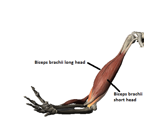
- The biceps brachii known as the biceps located in the anterior compartment of the arm.
- The biceps consists of two heads, the long and the short head, which arise proximally and joint to attach to the radius as a biceps tendon.
- Origin :
- Long head: Supra-glenoid tubercle of the scapula.
- Short head: Apex of the coracoid process of the scapula.
- Insertion :
- Radial tuberosity of the radius
- Deep fascia of the forearm as a bicipital aponeurosis.
- Nerve supply :
- Musculocutaneous nerve (C5-C6)
- Blood supply :
- Branch of brachial artery
- Action :
- Flexion and supination of the forearm at the elbow joint,
- Also, act as a flexor of the arm at the glenohumeral joint with the help of a long head.
- Clinical relation :
- 1)Pulley lesion is characterized by damage to the biceps pulley complex through which the long biceps tendon is no longer secured in the shoulder joint and thus slips out of the intertubercular sulcus.
- 2)Biceps tendinitis is an inflammation of the long biceps tendon often caused by bursitis or tendinitis involving the rotator cuff.
- 3)In severe cases the tendon can even tear apart completely known as biceps tendon rupture.
Triceps
- The triceps brachii is the muscle that runs down the back of the humerus.
- The triceps brachii gets its name because it contains three muscle ‘heads'( points of origin).
- These include the:
- 1)Medial head
- 2)Lateral head
- 3)Long head
- Origin:
- Long head: infra glenoid tubercle of the scapula
- Medial head: Inferior to radial groove on the posterior surface of the humerus.
- Lateral head: Superior to radial groove on the posterior surface of the humerus.
- Insertion:
- Olecranon of ulna and fascia of forearm
- Nerve supply:
- Radial nerve (C6 to C8)
- Blood supply:
- The deep brachial artery, Superior ulnar collateral artery.
- Action:
- Extension of the forearm at the elbow joint.
- Extension and adduction of the arm at the shoulder joint with the help of a long head.
- Clinical relation:
- The triceps reflex, elicited by hitting the triceps, is often used to test the function of the nerves of the arm.
- This tests spinal nerves C6 and C7, predominately C7
FAQs
What are the 3 main muscles in your shoulders?
The rotator cuff muscles, Deltoids, and Pectoral muscle are the main muscle group that supports the shoulder joint.
The four Rotator cuff muscles are
the supraspinatus
infraspinatus
teres minor
subscapularis.
What are the 9 muscles of the shoulder joint?
There are 9 muscles of the shoulder joint are:
Biceps brachii – long head
Triceps brachii – long head
Deltoid.
Supraspinatus.
Infraspinatus.
Teres minor.
Subscapularis.
Teres major.
How many muscle parts are in the shoulder?
Total is 8 muscles
This joint is supported by around 8 muscles in your shoulder. They provide it with strength, stability, and form. The muscles in your shoulders are skeletal muscles. Tendons connect them to the bones.
82 Comments